
The Ultimate Guide to GLP-1 vs “GLP-3” (Retatrutide): Molecular Mechanisms and Biohacking Protocols

Disclaimer: The information provided in this article is strictly for educational and informational purposes. The compounds discussed, including investigational triple hormone receptor agonists, are strictly for Laboratory Research Use Only and are not approved for human consumption. This content is intended for advanced scientific researchers and data analysts to understand molecular mechanisms and is not intended to diagnose, treat, or cure any biological condition.
Table of Contents
- Quick Answer: The Core Difference Between GLP-1 and GLP-3
- Understanding the Incretin System
- GLP-1 Receptor Agonists: The Current Gold Standard
- Enter “GLP-3”: The Rise of Triple Hormone Receptor Agonists
- Head-to-Head: GLP-1 vs GLP-3 Molecular Mechanisms
- Laboratory Synthesis and Clinical Data Analysis
- Advanced Biohacking Protocols: Transitioning
- Sourcing, Purity Standards, and Third-Party Testing
- Safety, Side Effects, and Managing Receptor Fatigue
- The Future of Metabolic Peptides
- Frequently Asked Questions (FAQs)
- Key Takeaways
Quick Answer: The Core Difference Between GLP-1 and GLP-3
When comparing GLP-1 vs GLP-3, the core difference lies in receptor targeting. GLP-1 agonists target a single metabolic pathway to regulate insulin and appetite. “GLP-3″—the biohacking term for investigational triple-agonists like retatrutide—simultaneously targets GLP-1, GIP, and glucagon receptors, synergistically driving unprecedented fat oxidation, appetite suppression, and increased resting metabolic rate.
Understanding the Incretin System: The Foundation of Metabolic Peptides
To fundamentally grasp the ongoing revolution in metabolic peptides and fully appreciate the leap from single receptor agonism to triple receptor agonism, we must first map the bioenergetic landscape of the human incretin system. The incretin effect describes the biological phenomenon where oral glucose ingestion elicits a vastly more robust insulin secretory response than an equivalent dose of intravenous glucose. This discrepancy is governed by gut-derived hormones called incretins, which act as nutrient sensors and metabolic orchestrators.
What is the GLP-1 Receptor?
Glucagon-Like Peptide-1 (GLP-1) is the most heavily researched incretin hormone to date. It is primarily synthesized and secreted by enteroendocrine L-cells located in the distal ileum and colon in response to nutrient ingestion (specifically lipids and carbohydrates).
At the molecular level, GLP-1 binds to the GLP-1 receptor (GLP-1R), which is a G-protein-coupled receptor (GPCR). When this binding occurs on the pancreatic beta cells, it initiates an intracellular signaling cascade involving the activation of adenylyl cyclase, a subsequent rise in cyclic AMP (cAMP), and the influx of intracellular calcium. This sequence strictly triggers glucose-dependent insulin secretion, meaning it only forces insulin release in the presence of elevated blood sugar, drastically minimizing the risk of iatrogenic hypoglycemia.
Beyond the pancreas, the GLP-1 receptor is highly expressed in the central nervous system (specifically the hypothalamus and hindbrain) and the gastrointestinal tract. Activation in the brain aggressively blunts orexigenic (appetite-stimulating) signaling, while activation in the gut delays gastric emptying. This two-pronged effect drastically reduces caloric intake, forming the bedrock of modern pharmaceutical weight management.
The Roles of GIP and Glucagon
While GLP-1 focuses heavily on satiety and insulin release, a comprehensive metabolic profile requires understanding two additional pathways: GIP and Glucagon.
GIP (Glucose-dependent Insulinotropic Polypeptide): Secreted by K-cells in the duodenum and jejunum, GIP is the sister hormone to GLP-1. It also acts as an incretin to stimulate insulin secretion. However, GIP receptors are uniquely and densely expressed in adipose (fat) tissue. Activation of the GIP receptor improves white adipose tissue blood flow and enhances lipid buffering capacity, effectively clearing circulating triglycerides and preventing ectopic fat deposition in the liver and skeletal muscle. Furthermore, GIP possesses potent anti-emetic properties, effectively mitigating the severe nausea often associated with heavy GLP-1 receptor activation.
Glucagon: Traditionally viewed purely as the counter-regulatory hormone to insulin, glucagon is secreted by pancreatic alpha cells to raise blood glucose via hepatic glycogenolysis (breaking down stored glycogen) and gluconeogenesis (synthesizing new glucose). For decades, researchers avoided glucagon agonism in metabolic therapies, fearing it would exacerbate hyperglycemia. However, modern biochemistry has revealed a paradox: when the glucagon receptor is activated in tandem with strong GLP-1 and GIP signaling, the hyperglycemic effects are entirely buffered by the incretin-driven insulin response. What remains is glucagon’s secondary, highly desirable function: the aggressive stimulation of lipolysis (fat breakdown) and a direct increase in hepatic energy expenditure and thermogenesis.
GLP-1 Receptor Agonists: The Current Gold Standard
Before looking toward the future of biohacking and advanced pharmacology, we must establish the baseline of the current gold standard. Single-agonist peptides have completely redefined the treatment of metabolic syndrome.
Mechanism of Action (From Semaglutide to Liraglutide)
The endogenous half-life of natural GLP-1 in the human body is roughly 1.5 to 2 minutes before it is rapidly cleaved and deactivated by the dipeptidyl peptidase-4 (DPP-4) enzyme. Therefore, early pharmacological engineering focused entirely on extending this half-life.
First-generation GLP-1s, such as exenatide (derived from the saliva of the Gila monster), offered marginal half-life extensions. Liraglutide advanced the field by attaching a C16 fatty acid chain to the peptide backbone, allowing it to bind to human serum albumin and evade rapid renal clearance, achieving a 13-hour half-life suitable for daily subcutaneous injection.
The breakthrough arrived with Semaglutide. Biochemists substituted an amino acid at position 8 (replacing alanine with alpha-aminoisobutyric acid) to completely shield the peptide from DPP-4 enzymatic degradation. Additionally, they attached a much larger C18 fatty diacid chain via a hydrophilic spacer. This robust molecular architecture extended the half-life to roughly 165 hours, allowing for once-weekly dosing. The clinical results—often averaging 15% total body weight loss over 68 weeks—solidified GLP-1 receptor agonists as the apex tools for metabolic control.
The Limitations and “The Plateau Effect”
Despite their profound efficacy, strict GLP-1 agonists carry inherent biological limitations. When researchers and advanced biohackers evaluate the landscape of metabolic interventions, the “plateau effect” is the most frequently cited hurdle.
The human body is an exquisite machine designed for homeostasis and survival. When subjected to prolonged caloric deficits driven by profound GLP-1-induced anorexia, the body initiates adaptive thermogenesis. It systematically downregulates thyroid function, reduces sympathetic nervous system tone, and lowers the resting metabolic rate (RMR) to conserve energy.
Simultaneously, chronic stimulation of the GLP-1 receptor leads to target-tissue tachyphylaxis (receptor downregulation). The receptors become desensitized to the peptide, requiring higher doses to achieve the same anorectic effect. In clinical literature, this presents as a hard stall in weight loss around months 12 to 16 of administration. The patient is eating less but no longer oxidizing adipose tissue because their metabolic engine has idled down to match the low caloric intake. To break this thermodynamic stall, a fundamentally different molecular mechanism is required.
Enter “GLP-3”: The Rise of Triple Hormone Receptor Agonists
The scientific pursuit of breaking the GLP-1 plateau led to the creation of unimolecular multi-agonists. In the gray market of research chemicals and the cutting edge of the biohacking community, a new term emerged to describe this evolution.
Why “GLP-3” is a Misnomer (But Crucial to Understand)
To maintain strict scientific accuracy, we must address the nomenclature. There is no naturally occurring hormone, receptor, or biological pathway known as “GLP-3”. Biologically, the incretin system does not contain a “Glucagon-Like Peptide 3.”
In the spheres of laboratory wholesale, underground peptide synthesis, and B2C biohacking forums, “GLP-3” has become the ubiquitous slang term for Retatrutide (developmental code LY3437943) and similar investigational triple hormone receptor agonists. The “3” signifies the simultaneous agonism of three distinct receptors: GLP-1, GIP, and Glucagon.
While technically a misnomer, understanding the “glp 1 vs glp 3” search intent is vital. When a researcher or biohacker searches for GLP-3, they are explicitly looking for data on triple-agonists and how they biochemically surpass single-pathway peptides.
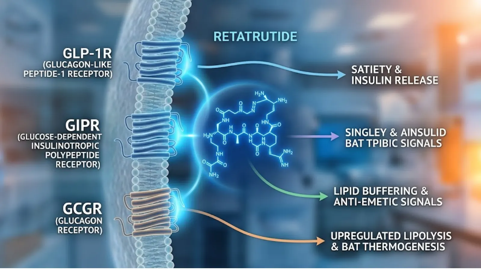
Fig 1: Conceptual diagram of Retatrutide simultaneously targeting GLP-1R, GIPR, and GCGR (Glucagon) pathways for synergistic metabolic drive.
Retatrutide’s Synergistic Triad
The biochemical engineering behind “GLP-3” (Retatrutide) is nothing short of a masterpiece in peptide synthesis. It is not a mixture of three different drugs; it is a single, heavily modified peptide backbone consisting of 39 amino acids.
The true genius lies in the titration of receptor agonism. It does not hit all three receptors equally. If it agonized the glucagon receptor too aggressively, it would cause severe hyperglycemia. Instead, the molecule is “biased.” It features highly potent GIP receptor agonism, moderate GLP-1 receptor agonism, and very carefully calibrated, weaker glucagon receptor agonism.
This triad works in perfect concert:
- GLP-1 suppresses appetite and slows gastric emptying.
- GIP profoundly sensitizes insulin, clears triglycerides, and directly mitigates the nausea caused by the GLP-1 action.
- Glucagon directly signals adipocytes (fat cells) to release stored lipids for fuel, bypassing the need for a caloric deficit to initiate lipolysis.
The result is not additive; it is synergistic. The clinical trial data for Retatrutide demonstrates an unprecedented 24.2% average body weight loss at 48 weeks—shattering the ceiling established by Semaglutide.
Head-to-Head: GLP-1 vs GLP-3 Molecular Mechanisms and Efficacy
To provide absolute clarity for both laboratory researchers and advanced biohacking cohorts, we must analyze the “glp 1 vs glp 3” debate across three highly specific, mechanism-driven vectors.
Fat Oxidation Rates Compared
With standard GLP-1 agonists, fat loss is a secondary downstream effect. The drug forces you to eat less, and the resulting caloric deficit eventually forces the body to mobilize fat stores for energy. It is an indirect mechanism of action for lipolysis.
“GLP-3” acts directly on adipose tissue. The inclusion of the glucagon receptor agonist directly upregulates hormone-sensitive lipase (HSL) within the white adipose tissue. HSL is the rate-limiting enzyme responsible for hydrolyzing stored triglycerides into free fatty acids and glycerol. Therefore, Retatrutide forces the cellular machinery to actively burn fat, independent of the caloric deficit. In laboratory murine models, triple agonists clear hepatic steatosis (fatty liver) with aggressive speed, fundamentally reversing lipotoxicity at the cellular level far faster than GLP-1 alone.
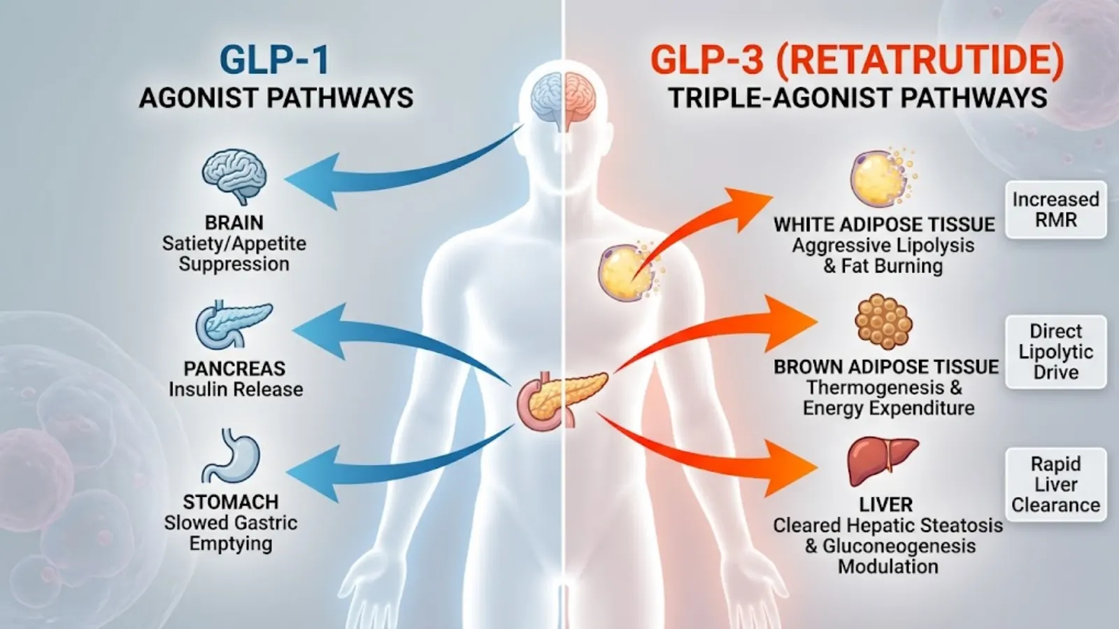
Fig 2: Physiological targeting map contrasting single-agonist GLP-1 pathways against the vastly expanded targets of a triple-agonist.
Impact on Resting Metabolic Rate (RMR)
This is arguably the most critical differentiator in the glp 1 vs glp 3 comparison. As previously noted, GLP-1s typically cause a drop in Resting Metabolic Rate. As weight is lost and caloric intake plummets, the body slows down.
“GLP-3” flips this thermodynamic equation. The glucagon component interacts with receptors in the liver and brown adipose tissue (BAT). BAT is a specialized form of fat rich in mitochondria that burns energy to produce heat (non-shivering thermogenesis). By activating BAT and increasing hepatic energy expenditure, Retatrutide successfully defends the metabolic rate against the expected adaptive thermogenesis. In short, while GLP-1 slows the metabolism down to protect energy stores, GLP-3 keeps the metabolic furnace burning hot despite massive caloric restriction.
Muscle Retention vs. Muscle Wasting
A major concern in the biohacking community regarding profound weight-loss peptides is sarcopenia (the loss of skeletal muscle tissue). Severe caloric restriction invariably leads to catabolism.
With standard GLP-1 therapies, muscle loss is common if the subject is not actively engaging in hypertrophy training and consuming adequate protein. Roughly 30-40% of the weight lost on early GLP-1 trials was lean mass.
The introduction of the glucagon receptor in GLP-3 complicates this picture. Glucagon is inherently a catabolic hormone; its job is to break down tissue for energy. Therefore, one might assume GLP-3 causes more muscle wasting. However, the profound insulin-sensitizing effects of the highly potent GIP component in Retatrutide create a nutrient-partitioning effect. It shuttles the limited available amino acids directly into muscle tissue while forcing the body to use lipids (liberated by glucagon) for systemic energy. While clinical data is still emerging on precise body composition ratios, early indicators suggest that if dietary protein is optimized, the lipolytic drive of GLP-3 spares lean tissue better than the generalized starvation mechanism of early GLP-1s.
Comparative Mechanisms: GLP-1 vs “GLP-3”
| Feature / Mechanism | Traditional GLP-1 (e.g., Semaglutide) | “GLP-3” / Triple Agonist (Retatrutide) |
|---|---|---|
| Receptor Targets | GLP-1R exclusively | GLP-1R, GIPR, and Glucagon Receptor |
| Primary Driver of Fat Loss | Severe caloric deficit via anorexia | Direct lipolysis via HSL upregulation + caloric deficit |
| Effect on Resting Metabolic Rate | Decreases (Adaptive Thermogenesis) | Increases / Sustains via BAT activation |
| Nausea / GI Distress | Moderate to Severe (dose-dependent) | Mild to Moderate (mitigated by GIP action) |
| Clearance of Hepatic Steatosis | Slow / Indirect | Rapid / Direct |
| Avg. Clinical Weight Loss (1 yr) | ~15% total body weight | ~24% total body weight (Phase 2 data) |
Laboratory Synthesis and Clinical Data Analysis
For both laboratory wholesalers sourcing active pharmaceutical ingredients (APIs) and researchers analyzing efficacy, the clinical milestones and synthesis pathways of these compounds are paramount. The jump from manufacturing a single-agonist peptide to a unimolecular triple-agonist is a monumental leap in biochemical engineering.
Current Clinical Trial Milestones for Retatrutide
When researchers evaluate the glp 1 vs glp 3 data, they look heavily at the Phase 2 clinical trial results published in the New England Journal of Medicine. In trials involving adults with obesity (BMI ≥ 30), retatrutide yielded a staggering mean weight reduction of 24.2% over 48 weeks at the highest dose (12 mg weekly).
More profoundly, 100% of the participants on the 8 mg and 12 mg doses achieved a weight reduction of at least 5%, and nearly half of the high-dose cohort lost over 25% of their total body weight. This velocity and magnitude of weight loss mimic the outcomes of bariatric surgery. Furthermore, researchers noted a complete normalization of hepatic fat in subjects with non-alcoholic fatty liver disease (NAFLD) within just 24 weeks, an effect heavily attributed to the glucagon receptor’s localized activity in the liver.
Synthesis Challenges and Molecular Stability
Synthesizing “GLP-3” (retatrutide) presents distinct challenges for laboratories. Unlike the 31-amino-acid structure of semaglutide, retatrutide relies on a 39-amino-acid backbone modeled heavily on the GIP sequence.
To achieve multi-receptor affinity without rapid enzymatic degradation, chemists employ Solid-Phase Peptide Synthesis (SPPS). They incorporate highly specific non-coded amino acids. Specifically, the substitution of alpha-aminoisobutyric acid (Aib) at position 2 is what protects the peptide chain from cleavage by the ubiquitous dipeptidyl peptidase-4 (DPP-4) enzyme.
Furthermore, to extend the pharmacokinetic half-life to allow for once-weekly dosing, a C20 fatty diacid moiety is conjugated to the lysine residue at position 17 via a hydrophilic linker. This lipid tail allows the molecule to bind tightly to circulating albumin in the bloodstream, preventing rapid renal clearance. For B2B synthesizers, ensuring the structural integrity of this lipid tail during the cleavage and global deprotection phases of SPPS is the most critical hurdle to achieving high-yield, stable batches of GLP-3.
Advanced Biohacking Protocols: Transitioning from GLP-1 to GLP-3
Disclaimer: The following protocols are theoretical frameworks discussed within advanced biohacking and experimental research communities. They do not constitute medical advice.
For researchers and biohackers utilizing these compounds in non-clinical settings to optimize body composition, transitioning from a standard GLP-1 to a highly potent “GLP-3” requires meticulous planning. The sheer metabolic force of a triple agonist can easily overwhelm a system adapted only to single-receptor agonism.
Dosing Strategies and Half-Life Considerations
Both semaglutide (GLP-1) and retatrutide (“GLP-3”) share a roughly similar pharmacokinetic half-life of approximately 6 to 7 days due to their albumin-binding fatty acid chains. However, receptor affinity and downstream biological responses differ vastly.
When biohackers hit the inevitable “GLP-1 plateau,” they often consider jumping immediately to a high dose of GLP-3. This is universally considered a poor protocol. Because the glucagon receptor is being introduced into the equation for the first time, sudden, high-dose administration can cause severe autonomic nervous system dysregulation, manifesting as hyperhidrosis (excessive sweating), severe tachycardia, and systemic anxiety.

Fig 3: Advanced biohacking flat lay illustrating the theoretical stacking of Retatrutide alongside secretagogues and amino acids for muscle preservation.
Micro-Dosing vs. Titration Protocols
The most highly regarded protocol for transitioning involves a mandatory “washout” period followed by micro-titration.
- The Washout: Ceasing GLP-1 administration for 14 to 21 days (roughly 2 to 3 half-lives). This allows the chronically down-regulated GLP-1 receptors to partially resensitize, and clears the metabolic slate.
- Micro-Titration: Initiating GLP-3 at a sub-clinical dose (e.g., 1 mg to 2 mg weekly). Biohackers often split this dose into bi-weekly subcutaneous injections (e.g., 1 mg every 3.5 days) to blunt the peak plasma concentration (Cmax) and minimize the acute introduction of glucagon-driven heart rate spikes. The dose is only titrated upward when the subject notes an adaptation to the resting energy expenditure increase.
Stacking Strategies for Muscle Preservation
Because the glucagon signaling in GLP-3 inherently drives catabolism to liberate energy, the preservation of lean skeletal muscle is the biohacker’s primary objective. The “glp 1 vs glp 3” dynamic means you are moving from a state of passive starvation to active tissue breakdown.
To counter this, advanced protocols heavily feature concurrent administration of muscle-sparing peptides and exogenous amino acids.
- Secretagogues: Compounds like CJC-1295 with Ipamorelin are frequently stacked to increase endogenous Growth Hormone (GH) pulses. Elevated GH directly opposes the protein-wasting effects of severe caloric deficits and synergizes with the fat oxidation pathways of the triple agonist.
- BPC-157: Often included to manage gastric inflammation and protect the mucosal lining of the gut during periods of drastically reduced gastric emptying.
- mTOR Activation: Biohackers strictly time their essential amino acid (EAA) intake—specifically leucine-heavy blends—around resistance training to aggressively spike the mTOR pathway, signaling the body to retain myofibrillar proteins despite the massive energy deficit.
Sourcing, Purity Standards, and Third-Party Testing
As the hype surrounding “GLP-3” reaches a fever pitch, the gray market for research peptides has become saturated with mislabeled, under-dosed, or completely counterfeit products.
Identifying Fake “GLP-3” in the Grey Market
Because synthesizing a 39-amino-acid peptide with a C20 fatty diacid chain is incredibly expensive and technically difficult, illicit laboratories frequently pass off cheaper, easily synthesized peptides as retatrutide. The most common scam is labeling standard semaglutide or Tirzepatide (a dual GIP/GLP-1 agonist) as GLP-3.
If a researcher administers what they believe is GLP-3 but notes no increase in thermogenesis, no resting heart rate elevation, and the exact same plateau effects they experienced on GLP-1, it is highly probable they have received a counterfeit single or dual agonist.

Fig 4: Laboratory analysis using HPLC and Mass Spectrometry to verify the purity and exact molecular weight of synthesized GLP-3 APIs.
The Importance of HPLC Testing and Mass Spectrometry
For both B2B wholesalers and B2C biohackers, relying on a Certificate of Analysis (COA) is non-negotiable. However, one must know how to read it. The gold standard for verifying peptide purity and identity is High-Performance Liquid Chromatography combined with Mass Spectrometry (HPLC-MS).
- HPLC (Purity): This process separates the compounds in the vial. A high-quality GLP-3 peptide should show a single, sharp, dominant peak on the chromatogram, indicating a purity of >99%. Multiple wide peaks indicate synthesis errors, truncated peptide chains, or heavy heavy metal contamination from the cleavage process.
- Mass Spectrometry (Identity): Purity is irrelevant if the molecule is wrong. Mass spec measures the exact molecular weight of the compound. Retatrutide has a highly specific molar mass (approximately 4731.3 g/mol). If the mass spec data shows the molecular weight of semaglutide (4113.6 g/mol), the product is fake, regardless of its 99% purity.
Lyophilized Powder Storage Best Practices
Research peptides are shipped as lyophilized (freeze-dried) powder. In this state, a GLP-3 peptide is relatively stable at room temperature for several weeks, provided it is kept out of direct ultraviolet (UV) light, which breaks down the peptide bonds.
However, once reconstituted with bacteriostatic water (water containing 0.9% benzyl alcohol to inhibit bacterial growth), the molecular clock begins ticking. The covalent bonds of the peptide chain are highly susceptible to hydrolysis in an aqueous solution. Reconstituted GLP-3 must be kept refrigerated at 2°C to 8°C (36°F to 46°F). Freezing reconstituted peptides is disastrous, as the formation of ice crystals mechanically shears the delicate peptide chains, rendering the compound biologically inert.
Safety, Side Effects, and Managing Receptor Fatigue
While the efficacy of triple agonists is undeniable, the side effect profile requires intense management. Overstimulating the incretin system and the glucagon pathway simultaneously carries unique physiological burdens.
Navigating Gastrointestinal Distress
Interestingly, upon initial administration, GLP-3 can sometimes produce less acute nausea than GLP-1. This is because the robust GIP receptor agonism acts as a powerful anti-emetic in the central nervous system, effectively buffering the nausea-inducing signals sent by the GLP-1 agonism. However, as the dose scales up, the profound slowing of gastric emptying will invariably lead to gastrointestinal distress, sulfur burps, and potential gastroparesis if dietary volume is not drastically reduced.
Mitigating Glucagon-Induced Tachycardia
This is the most critical safety differential in the glp 1 vs glp 3 paradigm. The glucagon receptor is present in the myocardium (heart muscle). Activation of this receptor has a positive chronotropic effect, meaning it directly increases the resting heart rate.
Clinical trials of retatrutide consistently show subjects experiencing an increase in resting heart rate of 5 to 10 beats per minute, which can peak at around 15 weeks before slowly declining. For biohackers, this can manifest as heart palpitations or decreased cardiovascular performance during high-intensity interval training (HIIT). Monitoring heart rate variability (HRV) via wearable bio-trackers is a mandatory protocol for anyone researching these compounds. If the resting heart rate spikes dangerously, the dosage must be immediately down-titrated.
Strategies for Resetting Peptide Tolerance
Continuous, uncycled use of any peptide leads to receptor tachyphylaxis (desensitization). The body’s homeostatic drive will eventually downregulate GLP-1, GIP, and Glucagon receptors to protect against what it perceives as an endless state of hyper-metabolic starvation.
Advanced researchers employ “drug holidays” to clear receptor fatigue. This involves systematically tapering off the GLP-3 compound over a 4-week period to avoid rebound hyperphagia (extreme, insatiable hunger). During the off-cycle, biohackers rely heavily on insulin-sensitizing agents (like berberine or metformin) and strict macronutrient control to maintain the new, lower body weight set point while the incretin receptors slowly return to baseline sensitivity.
The Future of Metabolic Peptides
The biochemical arms race does not end at triple agonists. As “GLP-3” moves closer to FDA approval for the masses, the vanguard of peptide research is already looking at combinations that completely bypass the incretin system’s limitations.
The next horizon involves pairing incretin agonists with Amylin analogs (such as Cagrilintide). Amylin is a hormone co-secreted with insulin that acts on entirely different brain centers to induce satiety and prevent the post-meal spike in glucagon.
Furthermore, because muscle loss remains the ultimate enemy of longevity, pharmaceutical companies are heavily investing in dual therapies: combining a metabolic fat-burner (like a GLP-1 or GLP-3) with a myostatin inhibitor or an activin type II receptor antagonist (like bimagrumab). This holy grail combination would forcefully strip adipose tissue via the incretin system while simultaneously forcing the body to lay down new skeletal muscle tissue, radically redefining human body composition without the need for extreme caloric restriction.
Frequently Asked Questions (FAQs)
Why is GLP-3 considered more effective for fat oxidation than GLP-1?
GLP-3 (retatrutide) includes a glucagon receptor agonist, which directly upregulates hormone-sensitive lipase in adipose tissue. Unlike GLP-1, which causes fat loss indirectly through starvation, GLP-3 directly forces fat cells to mobilize stored lipids for energy, drastically increasing total fat oxidation.
What are the stability differences between GLP-1 and GLP-3 research peptides?
Both GLP-1 and GLP-3 utilize lipid chains for half-life extension and require lyophilized powder storage. However, because GLP-3 has a longer 39-amino-acid chain, it is slightly more susceptible to hydrolysis and mechanical shearing once reconstituted, requiring strict cold-chain adherence (36°F to 46°F).
How do you safely transition from a GLP-1 to a GLP-3 peptide protocol?
To avoid severe autonomic dysregulation and extreme heart rate spikes from the newly introduced glucagon agonism, researchers suggest a 14 to 21-day washout period from GLP-1. This is followed by initiating GLP-3 at a sub-clinical micro-dose, slowly titrating upwards as tolerance builds.
Does the addition of the glucagon receptor in GLP-3 increase resting metabolic rate?
Yes. While traditional GLP-1 therapies usually lower your resting metabolic rate due to extreme caloric deficits (adaptive thermogenesis), the glucagon component in GLP-3 stimulates brown adipose tissue and increases hepatic energy expenditure, effectively keeping the metabolic rate elevated.
Which chromatography method is most accurate for testing GLP-3 peptide purity?
High-Performance Liquid Chromatography (HPLC) is the gold standard for testing GLP-3 purity, ensuring the vial contains only the target compound. However, HPLC must be paired with Mass Spectrometry to verify the exact molecular weight (approx. 4731.3 g/mol) to guarantee the identity of retatrutide.
Key Takeaways
- Mechanism Matters: The glp 1 vs glp 3 debate is defined by receptor targets. GLP-1 solely regulates appetite and insulin, whereas GLP-3 (retatrutide) synergistically targets GLP-1, GIP, and Glucagon for a compounded metabolic effect.
- Direct Lipolysis: The addition of the glucagon receptor in triple agonists allows the body to bypass the starvation requirement, actively instructing fat cells to release stored triglycerides.
- Metabolic Defense: GLP-3 combats the dreaded “weight loss plateau” by increasing resting energy expenditure, effectively overriding the body’s natural adaptive thermogenesis.
- Purity is Paramount: The gray market is flooded with counterfeit peptides. Advanced researchers must demand both HPLC (for purity) and Mass Spectrometry (for molecular identity) to avoid being sold cheaper, single-agonist peptides.
- Respect the Glucagon: The primary side-effect differentiator is cardiovascular. Glucagon agonism increases the resting heart rate, necessitating careful dosing, precise titration, and vigilant cardiovascular monitoring.